Clinical Report: Hypercementosis Progression and Acquired Concrescence Case
Overview
This case report documents a 62-year-old female with generalized hypercementosis progressing to acquired concrescence, particularly affecting mandibular teeth Nos. 25/26 and 29/30. The findings emphasize the importance of periodic dental examinations to identify such progression, which may complicate future dental extractions.
Background
Hypercementosis is characterized by excessive, non-neoplastic deposition of radicular cementum, often idiopathic or secondary to local or systemic factors. It can affect single or multiple teeth and is radiographically seen as cementum overgrowth within the periodontal ligament and lamina dura boundaries. While systemic conditions like Paget’s disease and acromegaly can cause generalized hypercementosis, local factors such as occlusal trauma also contribute. Concrescence, the fusion of adjacent teeth by cementum, is a rare complication that may arise from severe hypercementosis.
Data Highlights
Tooth Nos.
Periodontal Pocket Depths (mm)
Mobility
Radiographic Findings
29
Buccal: 3, Distal: 3
None
Grossly thickened, bulbous, blunted roots; concrescence with No. 30
30
Buccal: 2, Distal: 4
None
Grossly thickened, bulbous, blunted roots; concrescence with No. 29; incipient PDL space widening
Key Findings
Generalized hypercementosis observed in multiple quadrants, including mandibular and maxillary regions.
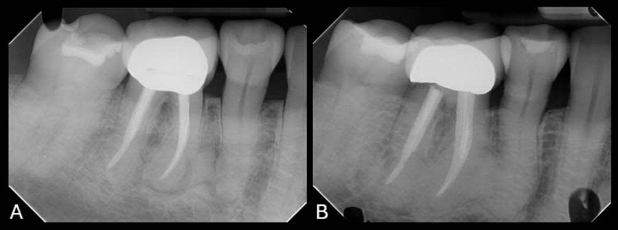
Progression of cementum deposition noted between 2018 and 2025, with development of concrescence in teeth Nos. 25/26 and 29/30.
Teeth Nos. 29 and 30 showed no mobility or percussion sensitivity despite severe root changes.
Patient had no systemic conditions commonly associated with hypercementosis, suggesting local occlusal trauma as the likely cause.
Radiographs revealed thickened, bulbous, and blunted roots with radiopaque cementum overgrowth confined within periodontal ligament boundaries.
Concrescence was acquired, indicated by fusion of cementum between adjacent teeth, complicating potential future extractions.
Clinical Implications
Clinicians should conduct regular radiographic and clinical evaluations to monitor hypercementosis progression, especially in patients with parafunctional habits or occlusal trauma. Awareness of possible concrescence is critical for treatment planning, as it may complicate extractions and require modified surgical approaches. Early detection can guide preventive strategies and minimize complications.
Conclusion
This case underscores the importance of periodic dental examinations to detect progressive hypercementosis and its rare complication, concrescence. Understanding these changes aids in anticipating treatment challenges and optimizing patient care.
Related Resources & Content
Gardner and Goldstein 1931 -- Definition of Hypercementosis
Midwestern University College of Dental Medicine-Arizona Case Report 2025 -- Hypercementosis Progression With Evidence of Acquired Concrescence