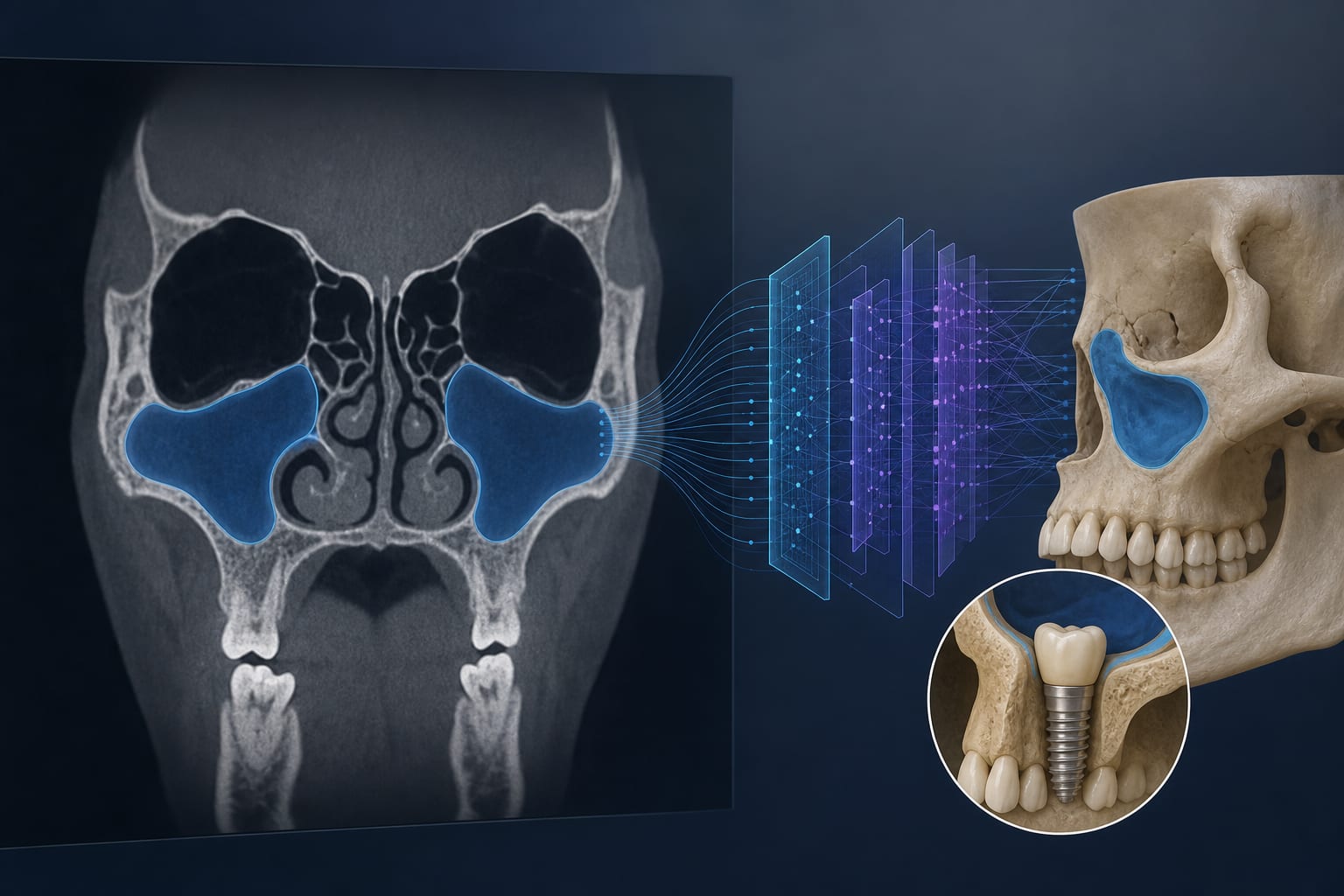
A Brief Review of AI Technology's Role in Maxillary Sinus Segmentation for Dental Applications
-
By
-
Jiayi Chen
-
May 20, 2026
Clinical Scorecard: A Brief Review of AI Technology's Role in Maxillary Sinus Segmentation for Dental Applications
At a Glance
| Category | Detail |
|---|
| Condition | Maxillary sinus segmentation |
| Key Mechanisms | Deep Learning (DL) for image analysis and segmentation |
| Target Population | Dental and ENT practitioners, junior physicians, graduates, and interns |
| Care Setting | Dental clinics and radiology departments |
Key Highlights
- DL enhances accuracy in identifying maxillary sinus boundaries on CT/CBCT images.
- Automated segmentation can save time and reduce bias in medical imaging.
- Integration of segmentation algorithms into radiology software is expected to improve medical education.
Guideline-Based Recommendations
Diagnosis
- Utilize DL models for accurate identification of maxillary sinus anatomy on imaging.
Management
- Incorporate automated segmentation in clinical practice to assist in procedures like sinus floor elevation.
Monitoring & Follow-up
- Apply advanced algorithms to monitor changes in maxillary sinus floor elevation post-surgery.
Risks
- Be aware of anatomical variations that may affect segmentation accuracy.
Patient & Prescribing Data
Patients requiring dental implants or treatment for maxillary sinus inflammation.
DL models can assist in evaluating the suitability of maxillary sinus for surgical interventions.
Clinical Best Practices
- Ensure adequate training sample sizes for DL models to avoid overfitting.
- Educate junior practitioners on the use of automated segmentation tools.
Related Resources & Content