Regenerative Materials in Site Preparation for Implant Placement: A Clinically Validated Histological Perspective
-
By
-
Robert A. Horowitz
-
Gregori M. Kurtzman, DDS
-
Hari S. Prasad BS
-
July 1, 2026
-
Clinical Scorecard: Regenerative Materials in Site Preparation for Implant Placement: A Clinically Validated Histological Perspective
At a Glance
| Category | Detail |
|---|
| Condition | Bone deficiencies in implant dentistry |
| Key Mechanisms | Integration of biologic mediators, osteoconductive scaffolds, and resorbable barrier membranes |
| Target Population | Patients requiring implant placement or prosthetic restoration |
| Care Setting | Periodontal and implant-related treatment planning |
Key Highlights
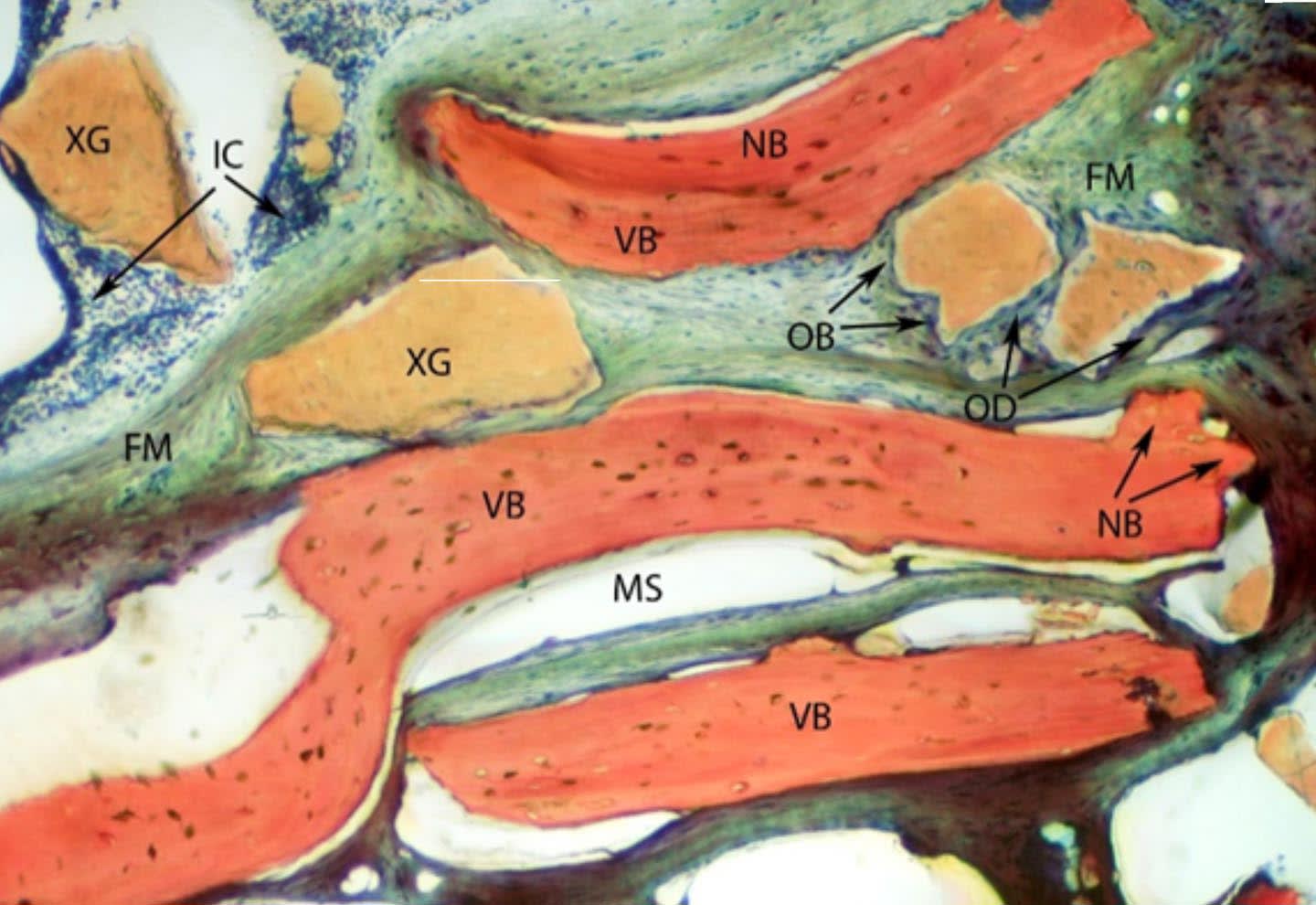
- Regenerative techniques preserve ridge volume and enhance implant stability
- Growth factor therapy accelerates vascular ingrowth and cellular migration
- Xenografts provide long-term volumetric stability; allografts show active remodeling
- Successful outcomes depend on wound stability and biologic containment
- Combination of biologic stimulation and mechanical stability is essential
Guideline-Based Recommendations
Diagnosis
- Assess bone deficiencies and periodontal support prior to implant placement
Management
- Utilize growth factor-enhanced matrices and osteoconductive scaffolds for regeneration
Monitoring & Follow-up
- Evaluate healing and integration of graft materials post-implant placement
Risks
- Potential for inadequate remodeling and soft tissue encroachment without proper techniques
Patient & Prescribing Data
Individuals with tooth extraction or periodontal disease
Use of rhPDGF-BB with scaffolds can enhance regenerative outcomes
Clinical Best Practices
- Maintain wound stability during regenerative procedures
- Employ barrier membranes to exclude epithelium when indicated
- Combine biologic mediators with mechanical scaffolds for optimal results
Related Resources & Content